
Database Accession: DI1100088
Name: TIR1 ubiquitin ligase in complex with Auxin-responsive protein IAA7
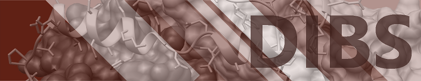
PDB ID: 2p1q
Experimental method: X-ray (1.91 Å)
Source organism: Arabidopsis thaliana
Proof of disorder:
Primary publication of the structure:
Tan X, Calderon-Villalobos LI, Sharon M, Zheng C, Robinson CV, Estelle M, Zheng N
Mechanism of auxin perception by the TIR1 ubiquitin ligase.
(2007) Nature 446: 640-5
PMID: 17410169
Abstract:
Auxin is a pivotal plant hormone that controls many aspects of plant growth and development. Perceived by a small family of F-box proteins including transport inhibitor response 1 (TIR1), auxin regulates gene expression by promoting SCF ubiquitin-ligase-catalysed degradation of the Aux/IAA transcription repressors, but how the TIR1 F-box protein senses and becomes activated by auxin remains unclear. Here we present the crystal structures of the Arabidopsis TIR1-ASK1 complex, free and in complexes with three different auxin compounds and an Aux/IAA substrate peptide. These structures show that the leucine-rich repeat domain of TIR1 contains an unexpected inositol hexakisphosphate co-factor and recognizes auxin and the Aux/IAA polypeptide substrate through a single surface pocket. Anchored to the base of the TIR1 pocket, auxin binds to a partially promiscuous site, which can also accommodate various auxin analogues. Docked on top of auxin, the Aux/IAA substrate peptide occupies the rest of the TIR1 pocket and completely encloses the hormone-binding site. By filling in a hydrophobic cavity at the protein interface, auxin enhances the TIR1-substrate interactions by acting as a 'molecular glue'. Our results establish the first structural model of a plant hormone receptor.
 Annotations from the GeneOntology database. Only terms that fit at least two of the interacting proteins are shown.
 Molecular function:
Biological process:
auxin-activated signaling pathway A series of molecular signals generated by the binding of the plant hormone auxin to a receptor, and ending with modulation of a downstream cellular process, e.g. transcription.
Cellular component:
Structural annotations of the participating protein chains.Entry contents: 2 distinct polypeptide molecules
Chains: C, B
Notes: Chain A was removed as chains C and B highlight the biologically relevant interaction.
Name: Auxin-responsive protein IAA7
Source organism: Arabidopsis thaliana
Length: 13 residues
Sequence:Sequence according to PDB SEQRESQVVGWPPVRNYRK
UniProtKB AC: Q38825 (positions: 82-94) Coverage: 5.3%
UniRef90 AC: UniRef90_Q38825 (positions: 82-94)
Name: Protein TRANSPORT INHIBITOR RESPONSE 1
Source organism: Arabidopsis thaliana
Length: 594 residues
Sequence:Sequence according to PDB SEQRESMQKRIALSFPEEVLEHVFSFIQLDKDRNSVSLVCKSWYEIERWCRRKVFIGNCYAVSPATVIRRFPKVRSVELKGKPHFADFNLVPDGWGGYVYPWIEAMSSSYTWLEEIRLKRMVVTDDCLELIAKSFKNFKVLVLSSCEGFSTDGLAAIAATCRNLKELDLRESDVDDVSGHWLSHFPDTYTSLVSLNISCLASEVSFSALERLVTRCPNLKSLKLNRAVPLEKLATLLQRAPQLEELGTGGYTAEVRPDVYSGLSVALSGCKELRCLSGFWDAVPAYLPAVYSVCSRLTTLNLSYATVQSYDLVKLLCQCPKLQRLWVLDYIEDAGLEVLASTCKDLRELRVFPSEPFVMEPNVALTEQGLVSVSMGCPKLESVLYFCRQMTNAALITIARNRPNMTRFRLCIIEPKAPDYLTLEPLDIGFGAIVEHCKDLRRLSLSGLLTDKVFEYIGTYAKKMEMLSVAFAGDSDLGMHHVLSGCDSLRKLEIRDCPFGDKALLANASKLETMRSLWMSSCSVSFGACKLLGQKMPKLNVEVIDERGAPDSRPESCPVERVFIYRTVAGPRFDMPGFVWNMDQDSTMRFSRQIITTNGL
UniProtKB AC: Q570C0 (positions: 1-594) Coverage: 100%
UniRef90 AC: UniRef90_Q570C0 (positions: 1-594)
Evidence demonstrating that the participating proteins are unstructured prior to the interaction and their folding is coupled to binding. Chain C:
The protein region involved in the interaction contains a known functional linear motif (DEG_SCF_TIR1_1).
Chain B:
The TIR1 domain involved in the interaction is known to adopt a stable structure in isolation. A solved structure of the domain without bound ligand peptides is represented by PDB ID 2p1p.
Structures from the PDB that contain the same number of proteins, and the proteins from the two structures show a sufficient degree of pairwise similarity, i.e. they belong to the same UniRef90 cluster (the full proteins exhibit at least 90% sequence identity) and convey roughly the same region to their respective interactions (the two regions from the two proteins share a minimum of 70% overlap). The structure can be rotated by left click and hold anywhere on the structure. Representation options can be edited by right clicking on the structure window.
Download our modified structure (.pdb)
